So ein ding: SDU can provide you with a very close look at the bumblebee
A whole new world opens up before you when you look through a microscope. At SDU, we have some very unique microscopes, which not only help our researchers but also companies, in connection with analysing and characterising materials and surfaces, which gives completely new insights into why a product may fail.
A bumblebee, gold, a virus and salt and pepper. Perhaps not the most obvious photo opportunities, but seen under a microscope, a new world opens up before us - a micro world where the physics is different and where other laws apply.
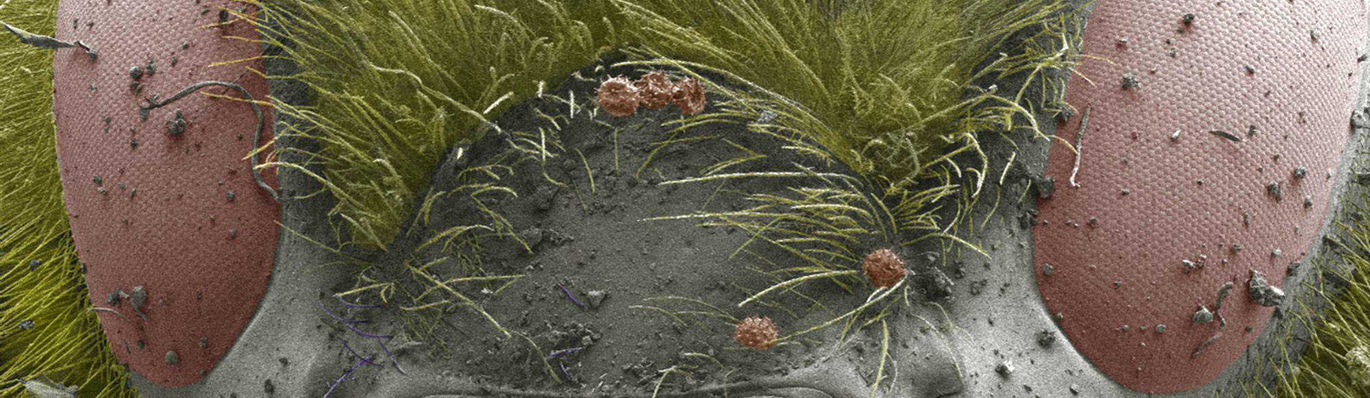
In this article, we get very close to our unique microscopes and the tasks that SDU can solve with them. At the same time, we dive into a world where a small creature like a bumblebee appears gigantic, and where you can not only look it in its eye but even see how the pollen is attached to its cilia.
Arkadiusz Goszczak from the ‘Center for Materials Analysis and Characterization’ (C:MAC red.) has taken many beautiful and fascinating pictures through some of the fantastic microscopes found at SDU.
- C:MAC is an umbrella organisation at SDU, where we have gathered a number of the unique facilities that we have. It ranges from microscopes to cleanrooms, but common to it all is that it can help companies and researchers analyse and characterise materials. With C:MAC, you get a quick overview of which services and expertise we have in this field, says Arkadiusz Goszczak.
Let's return to the bumblebee. In fact, it has a common denominator with the microscope with which the image of it is taken. It is said that bumblebees should not be able to fly, but they just do not know it themselves. The statement supposedly originated from Germany in the 30s, where a researcher in aerodynamics, concluded that a bee should not be able to fly according to his calculations. That should not be possible. The aerodynamicist had not taken into account that the bumblebee’s wings are rough and pliable in his calculations, which makes a colossal difference.
Until approximately 20-25 years ago, it was also widely accepted that in a light microscope one cannot distinguish structures that are smaller than approximately half the wavelength of the light used to study your sample. It was formulated as a physical law by Ernst Abbe in 1873 and is due to the refraction of light.
Today, however, it is possible to see details that are a thousand times smaller than the wavelength of light using nanoscopy. By studying the smallest building blocks - molecules and atoms - it is possible to understand how the world is connected. The impossible has become possible.
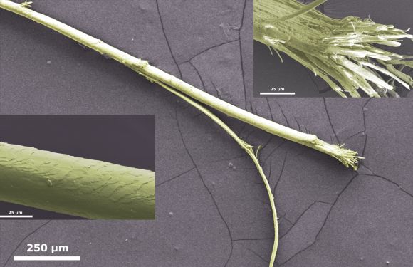
One of the smallest things we can see with the naked eye is the thickness of a strand of hair. It looks like this Under SDU's Specular Microscope CEM-530:
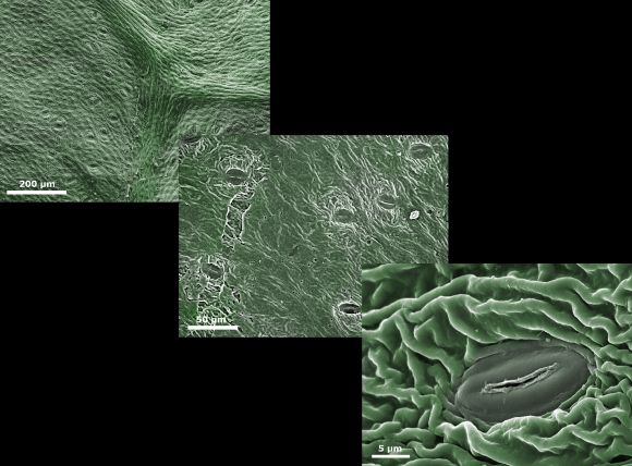
If we go very close to a leaf, we can also see that plants use microscopic 'mouths':
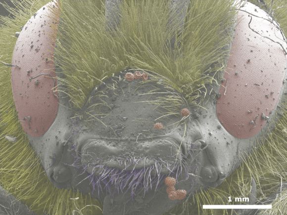
SDU's Specular Microscope CEM-530 is a fantastic instrument. The advantage of a scanning electron microscope is that we can look at larger samples, which in this context refers to, for example, a whole bumblebee. However, there are also disadvantages to an electron microscope. The sample must first be dead. The electron beams are highly energetic and can damage the material.
- However, we get around this by protecting the material with a coating, but I would like to emphasise in this context that no animal suffered damage in connection with the image of the bumblebee. It was dead in advance. Though I did steal the leaf from my mother's flower bed, so here an excuse might be in order. "Sorry Mom," laughs Arkadiusz Goszczak.
With the ORION helium ion microscope, you can not only see the surface of the sample but also easily see through tissues, muscles, bacteria. The microscope is in many ways similar to the CEM-530 microscope, but instead of electrons, it uses focused ion beams to generate both images and create nanostructures.
What can be seen here are structures, produced by Jacek Fiutowski, Head of the Nanophotonics group at NanoSYD, by using the ORION helium ion microscope.
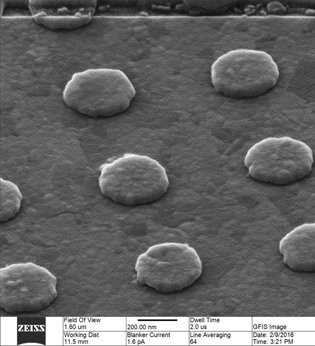 The gold dots are fabricated using Electron Beam Lithography.
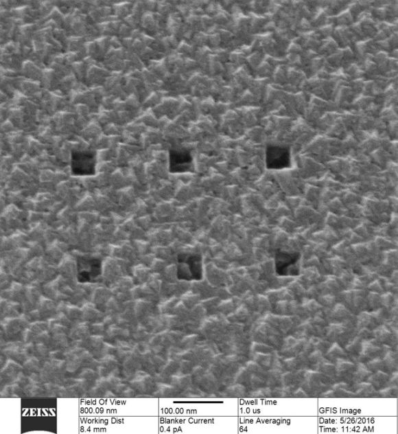
Here you can see square patterns that are only about 50nm wide and fabricated by using the ion milling option that the ORION helium ion microscope makes possible.
Below you can see a picture of adenovirus, a virus that causes respiratory diseases, gastrointestinal diseases, and eye infections.
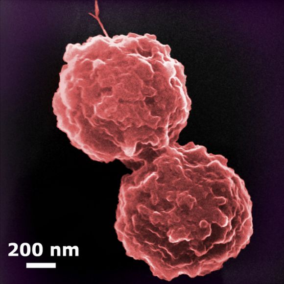
- It is important for understanding processes and mechanisms if we are to develop new drugs or develop vaccines. With this, it is important to be able to study tests on a level that is as detailed as possible, says Arkadiusz Goszczak.
You can read more about C:MAC - Center for Materials Analysis and Characterization here.
We have brand new state-of-the-art equipment, we have the equipment you will not find anywhere else, and then we have equipment that is just really, really cool and makes for a good story.
SDU has thingamabobs, doohickies, whatchamacallits and thousands of electronic devices that you will not find at home or in your company. And the cool thing is, you can make use of them. SDU kindly provides knowledge and equipment if we can find a joint project.
In this edition of the article series, the fantastic microscopes we have at SDU are in focus.
In this article, we get very close to our unique microscopes and the tasks that SDU can solve with them. At the same time, we dive into a world where a small creature like a bumblebee appears gigantic, and where you can not only look it in its eye but even see how the pollen is attached to its cilia.
Arkadiusz Goszczak from the ‘Center for Materials Analysis and Characterization’ (C:MAC red.) has taken many beautiful and fascinating pictures through some of the fantastic microscopes found at SDU.
- C:MAC is an umbrella organisation at SDU, where we have gathered a number of the unique facilities that we have. It ranges from microscopes to cleanrooms, but common to it all is that it can help companies and researchers analyse and characterise materials. With C:MAC, you get a quick overview of which services and expertise we have in this field, says Arkadiusz Goszczak.
Let's return to the bumblebee. In fact, it has a common denominator with the microscope with which the image of it is taken. It is said that bumblebees should not be able to fly, but they just do not know it themselves. The statement supposedly originated from Germany in the 30s, where a researcher in aerodynamics, concluded that a bee should not be able to fly according to his calculations. That should not be possible. The aerodynamicist had not taken into account that the bumblebee’s wings are rough and pliable in his calculations, which makes a colossal difference.
Until approximately 20-25 years ago, it was also widely accepted that in a light microscope one cannot distinguish structures that are smaller than approximately half the wavelength of the light used to study your sample. It was formulated as a physical law by Ernst Abbe in 1873 and is due to the refraction of light.
Today, however, it is possible to see details that are a thousand times smaller than the wavelength of light using nanoscopy. By studying the smallest building blocks - molecules and atoms - it is possible to understand how the world is connected. The impossible has become possible.
Electron microscope
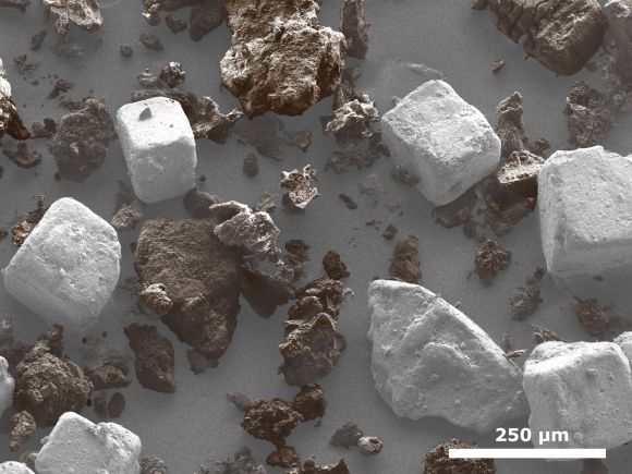
At SDU, Arkadiusz Goszczak uses an electron microscope to study things at nano-level. Electrons function in many ways the same as light, but have extremely short wavelengths, giving a high resolution. It makes it possible to see details like pollen in the bumblebee's face. Or, as shown below, salt and pepper:One of the smallest things we can see with the naked eye is the thickness of a strand of hair. It looks like this Under SDU's Specular Microscope CEM-530:
If we go very close to a leaf, we can also see that plants use microscopic 'mouths':
SDU's Specular Microscope CEM-530 is a fantastic instrument. The advantage of a scanning electron microscope is that we can look at larger samples, which in this context refers to, for example, a whole bumblebee. However, there are also disadvantages to an electron microscope. The sample must first be dead. The electron beams are highly energetic and can damage the material.
- However, we get around this by protecting the material with a coating, but I would like to emphasise in this context that no animal suffered damage in connection with the image of the bumblebee. It was dead in advance. Though I did steal the leaf from my mother's flower bed, so here an excuse might be in order. "Sorry Mom," laughs Arkadiusz Goszczak.
The crown jewel of the collection
Where SDU's Specular Microscope CEM-530's capabilities run short, another of SDU's microscopes steps in. The Mads Clausen Institute is the first in all of Scandinavia to have an ORION helium ion microscope installed.With the ORION helium ion microscope, you can not only see the surface of the sample but also easily see through tissues, muscles, bacteria. The microscope is in many ways similar to the CEM-530 microscope, but instead of electrons, it uses focused ion beams to generate both images and create nanostructures.
What can be seen here are structures, produced by Jacek Fiutowski, Head of the Nanophotonics group at NanoSYD, by using the ORION helium ion microscope.
The gold dots are fabricated using Electron Beam Lithography.Here you can see square patterns that are only about 50nm wide and fabricated by using the ion milling option that the ORION helium ion microscope makes possible.
Below you can see a picture of adenovirus, a virus that causes respiratory diseases, gastrointestinal diseases, and eye infections.
- It is important for understanding processes and mechanisms if we are to develop new drugs or develop vaccines. With this, it is important to be able to study tests on a level that is as detailed as possible, says Arkadiusz Goszczak.
You can read more about C:MAC - Center for Materials Analysis and Characterization here.
So ein Ding
We want to put all the fantastic equipment we have at the Faculty of Engineering into focus. That's why we are bringing the article series So ein Ding, the headline of which we have stolen from a popular DR program, that deals with electronic gadgets. Because when it comes to electronic equipment, few who can compete with SDU.We have brand new state-of-the-art equipment, we have the equipment you will not find anywhere else, and then we have equipment that is just really, really cool and makes for a good story.
SDU has thingamabobs, doohickies, whatchamacallits and thousands of electronic devices that you will not find at home or in your company. And the cool thing is, you can make use of them. SDU kindly provides knowledge and equipment if we can find a joint project.
In this edition of the article series, the fantastic microscopes we have at SDU are in focus.