Biomedical imaging
Structural characterisation of soft-matter and biological tissue samples
Keywords and applications
Keywords
Ultrahigh resolution imaging; biomimetics; nanoparticles
Applications
Biomimetics; implants; nanotoxicity; subcellular structure characterisation
Profile
In the field of biomedical imaging we are interested in the structural characterisation of soft-matter and biological tissue samples.
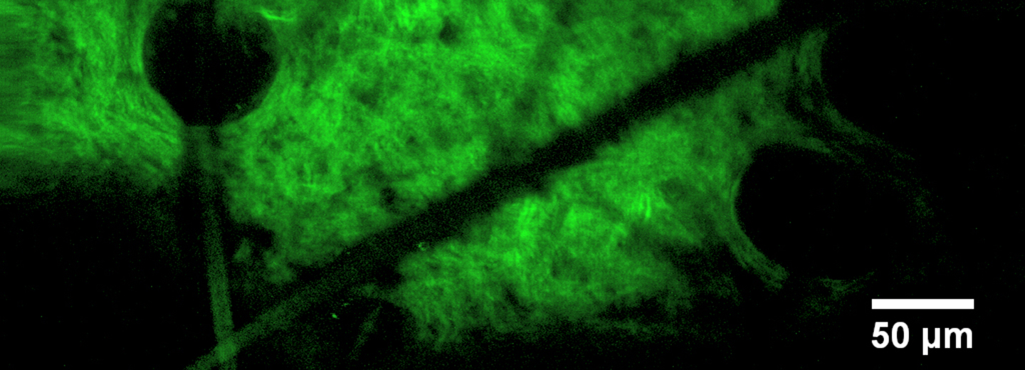
For example, in our Celltom project, we investigate the sub-cellular structure of animal and human tissue to improve the speed and reliability of optical diagnosis tools. For this, we use a correlative microscopy approach that combines ultrahigh resolution imaging of tissue blocks from biopsies with optical microscopy and tomography.
At our institutes, we conduct our research with a Helium ion microscope to avoid typical problems such as sectioning of tissue samples and to compensate for charging effects that lower the resolution in electron microscopy. In addition, an X-ray nanotomograph allows 3D imaging of entire tissue blocks, e.g. to identify relevant spots or segmentation.
In another project, we determined the fine structure of a very prominent biomaterial, i.e. spider silk, to facilitate advances in biomimetics.
In the future, we will extend our research activities towards nanoparticle characterisation in biomaterials and food products to study related health and safety issues.
Services
- Ultrahigh resolution imaging of dried and fixated biomaterials
- High resolution X-ray tomography of small and large biological samples
- Correlative optical and ion microscopy
Equipment
- Optical microscopes (bright-field, dark-field, fluorescence, fluorescence lifetime)
- Helium-ion microscope (< 10 nm resolution)
- X-ray nanotomograph (< 1 µm resolution).
Contact
Dr. Till Leißner
Associate Professor
SDU NanoSYD
The Mads Clausen Institute
T +45 6550 8378
till@mci.sdu.dk