Researchers want to map your cells. All 37 trillion of them!
We are gradually learning more about the cells of the human body than any researcher has ever dared to dream of. But what’s the purpose?
The mere thought of what one plunges into when deciding to research cells is mind-blowing: The human body contains 37 trillion cells, plus/minus a few billion.
Despite the fact that they contain the same DNA, each cell behaves differently, depending on how they are affected by the body and each other.
As if this wasn’t enough, they can also switch between different stages, depending on the challenges they face.
New ones are cropping up
That means a liver cell isn’t just a liver cell, and a skin cell isn’t just a skin cell. That’s the opposite of what researchers believed 50 years ago.
Back then, it was also estimated that there were around 200 different cell types, but increasingly advanced technology has allowed us to study the mechanisms in individual cells, and this has led to the discovery of many new cell types.
The philosophical question of cells
Throughout our lives, our cells are constantly being replaced. Every single cell in our body eventually dies and is replaced by a new one. For example, the intestinal cells are replaced in a few days, skin cells in a month's time and liver cells last for about six weeks. Philosophers argue that one may look upon the human body as the ship of Theseus. He was a Greek king whose ship was preserved long after his death. The ship was made of wood, and as the parts rotted, they were replaced with new wood. This prompted the Greek philosopher Plutarch to ask: Is this still the original ship? And if not, at which point did it cease to be the ship of Theseus? We can ask ourselves the same question: Does a cell remain the same once it has been replaced? And does a human remain the same when all of its cells have been replaced?
And new ones keep cropping up.
With that many different cell types, which can even switch between different stages, will we ever be able to fully comprehend how the human body works?
– Even though there are so many types of cells, and even though they interact differently, we have a good overview of the basic, general mechanisms for how, for example, a liver works.
– But it's like doing space research: We have an overview and qualified assumptions about what is out there – but we are also constantly discovering new planets and particles that lead to new knowledge and insight, says Kedar Natarajan.
Embryonic stem cells
He is an Assistant Professor and Head of Research at the Danish Institute of Advanced Science and the Department of Biochemistry and Molecular Biology.
His specialty is embryonic stem cells – which processes are in play when a fertilised egg begins to divide and give rise to all of the different cell types needed to build an organism?
He is also working on developing and improving techniques to find all of the currently undiscovered cell types and cell stages whose existence researchers are certain of.
An atlas of cells
The exploration and mapping of cell types and stages is a gigantic project that cannot be carried out by individual research groups. Large international collaborations have therefore been established, such as The Human Cell Atlas, which was founded in 2016.
The inventor behind this ambitious project is Kedar Natarajan's former research consultant, Professor Sarah Teichmann of the Wellcome Sanger Institute in England.
She was fascinated by the huge prospects of one day having identified all types of cells and their myriads of varying stages in the human organism. This will enable researchers to understand and treat diseases to an unprecedented extent.
”It's amazing to see so many different kinds of expertise contribute to this gigantic task
In addition to The Human Atlas, Kedar Natarajan's research group also contributes to the pan-European research effort The LifeTime Initiative, which focuses in particular on developing new technologies for single-cell research.
– It's amazing to see so many different kinds of expertise contribute to this gigantic task: Researchers from many fields, including biology, physiology, epidemiology, chemistry, biophysics, data science and mathematics have joined forces and are investing an insane amount of resources into this task, says Kedar Natarajan.
Breakthrough for malaria
The breakthroughs have already begun to show. One example is malaria.
Last year, Mara Lawniczak's research group at the Sanger Institute, with contributions from Kedar Natarajan, among others, announced the discovery of previously unknown types of cells responsible for the development of malaria.
However, since it’s only occasionally and under certain conditions that these cells actually trigger malaria, another interesting aspect is that the researchers were also able to pinpoint the specific conditions in which the cells became dangerous.
The really dangerous cancer cell
Kedar Natarajan and his colleagues at SDU are currently studying a gene found in most cells. Under normal conditions, this gene is harmless, but it can change, and in doing so, the cell becomes dangerous. Really dangerous, because the cell is now changing into a cancer cell.
– This gene is responsible for the development of most forms of cancer. Therefore, the fact that we’ve found the cell type that activates the gene and triggers the process that turns an otherwise healthy cell into a cancer cell could prove to be a huge breakthrough, says Kedar Natarajan.
This knowledge allows us to try to prevent the cell type from initiating the molecular chain reaction that leads to the development of all forms of cancer.
Too many cells in the blender
How can it be that it is only now that researchers have discovered that there are many more than 200 types of cells and that they can shape-shift into something almost unrecognisable?
It’s all a matter of perspective. 50 years ago, it made sense to assume that all cells in, for example, an organ such as the liver were more or less identical – or at least so similar that is was safe to assume that they behave in the same manner and react identically to influences.
Consequently, the characteristics of a liver cell were defined by, broadly speaking, blending a liver and then studying some of the cells that made up the blended mass to define what constitutes a liver cell.
”In the case of liver cells, we now know that there isn’t just one type. So far, we have defined 50 different types of liver cells.
What was not known at the time was that it was equivalent to emptying a fruit bowl into a blender: Sure, you get ‘fruit cells’ out of it, but you lose the level of detail and miss the fact that it originally contained many different cells from kiwis, bananas, apples etc.
– In the case of liver cells, we now know that there isn’t just one type. So far, we have defined 50 different types of liver cells, says Kedar Natarajan.
What happens once we’ve mapped them all?
This revolutionary insight came when advanced technology made it possible to study individual cells to an unprecedented extent. Today, single-cell research is a rapidly developing field with high-flying ambitions.
In addition to The Human Atlas, other international collaborations are constantly working to analyse single cells to find new types of cells and new cell stages, which can contribute to our understanding of our organism and how to cure it when we become ill.
Contributions are also collected in e.g. Pediatric Atlas, Lung Atlas, Brain Atlas, Liver Atlas etc.
Are we home and dry once we’ve mapped every single cell type and stage?
Further and deeper
– We will have solved that specific task. But then new ones will crop up. At that stage, we can start to look even deeper and, for example, address the individual molecules. Man's curiosity and desire for knowledge never stops, and therefore research never stops, either. We will constantly be searching further and deeper, Kedar Natarajan predicts.
What is being studied in each cell?
All cells contain genetic material, DNA and RNA. Genes can be activated or deactivated, and both conditions result in a specific cell behaviour. The RNA sequencing technique allows us to scan the genetic material of cells, and if we discover an undesirable activation which, for example, leads to the development of cancer, we can seek to de-activate that specific gene and thus prevent the cancer from starting in the first place. At SDU, this research is being carried out by, among others, the Functional Genomics and Metabolism research group.
Each cell contains approx. 10,000 different kinds of proteins and each type of protein is found in varying amounts, depending on the developmental state of the cell and its surrounding environment. Thus, there are more than 10 billion protein molecules in a cell, so developing new techniques that can analyse many different proteins simultaneously is paramount. This field of research is called proteomics. One of the major breakthroughs in proteomics, for example, is the identification of a number of proteins that play a role in stem cell differentiation; they affect what a stem cell develops into. By knowing them, it is possible to monitor and control the development of a stem cell and decide whether it should become a liver cell or a skin cell. At SDU, this is being studied at, among other places, the Protein Research Group at the Department of Biochemistry and Molecular Biology.
A myriad of biological processes take place inside a living cell, allowing the cell to metabolise nutrients, alter its interactions with neighbouring cells or initiate cell division. Using advanced microscopy equipment, we can observe these processes by looking at individual living cells, and thereby see and measure the changes inside the cell. At SDU, this is being studied at, among other places, the Danish Molecular Biomedical Imaging Center.
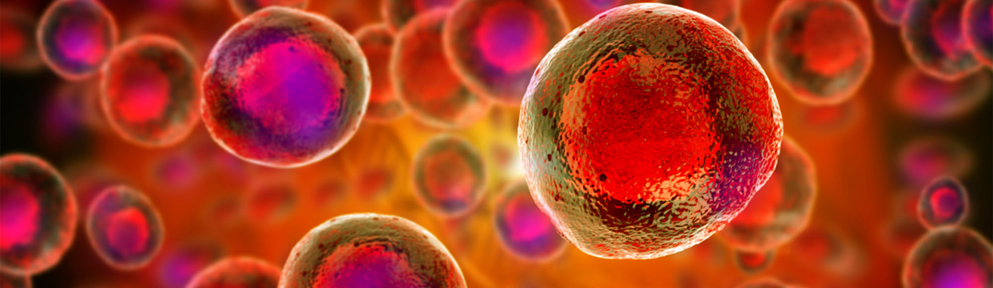
Top photo: Giovanni Cancemi for Adobe Stock
Meet the researcher
Kedar Natarajan is an assistant professor at the Department of Biochemistry and Molecular Biology. His research is supported by, among others, Villumfonden and Novo Nordisk Fonden.