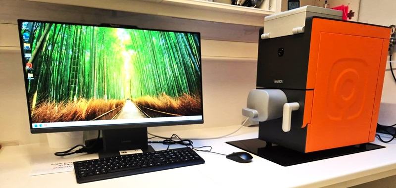
SRS Microscope

BondXplorer™ stimulated Raman scattering microscopy

Introduction:

The BondXplorer™ is a versatile, label-free microscope that uses Raman-based imaging to generate chemically specific pictures of tissues, cells, and materials. By combining stimulated Raman scattering (SRS) with multiphoton fluorescence and second harmonic generation (SHG), it reveals both chemical composition and structural details — all without staining or labels. Researchers can also record full spectral information, making it possible to identify and compare different molecules directly in the sample.

Applications:

·  Label-free tissue imaging – rapid virtual histology without dyes

·  Cancer vs. healthy tissue – chemical contrast for diagnostics and research

·  Spectral recording – identify and map molecules in situ

·  Drug delivery – track how compounds move in tissues and cells

·  Lipid transport in cells – visualize protein- and lipid-rich domains

·  Live enzyme activity – monitor metabolic processes in real time

·  Food and material samples – non-invasive chemical and structural imaging


Specifications

Imaging modalities

  • SRS (Stimulated Raman Scattering) – two simultaneous channels separated by 100 cm⁻¹
  • SRS spectral recording – capture and analyze full Raman spectra directly from the sample
  • CARS (Coherent Anti-Stokes Raman Scattering) – complementary vibrational imaging
  • Multiphoton fluorescence (2P/2PEF) – label-free or dye-based multiphoton imaging
  • SHG (Second Harmonic Generation) – structural imaging of ordered molecules (e.g., collagen)

Laser

  • APE Delta Emerald dual-color picosecond laser
  • Tunable across 350–3800 cm⁻¹ Raman range
  • Enables dual-SRS and nonlinear excitation for multiphoton and SHG

Objective

  • Nikon CFI Apo LWD 20× water-immersion multiphoton
  • NA 1.0, WD 2 mm – designed for deep tissue imaging with high near-IR transmission

Detection

  • 2 × balanced detectors for SRS channels
  • 3 × PMTs for SHG, multiphoton fluorescence, and CARS

Sample types

  • Compatible with glass slides, chambered coverslips, and culture dishes
  • Works with fixed or live biological tissues, organoids, food, and material samples

Imaging modes

  • 2D chemical and structural maps
  • Spectral mapping for identifying molecules in situ
  • Z-stacks for 3D volumes
  • Mosaic imaging for large-area scans (mm² scale)

Funding

National research infrastructure NanoChem


Figure: Label-free imaging of human skin using the BondXplorer™. Individual channels show SRS signals from CH (2939 cm¹, magenta) and CH (2856 cm¹, green), second harmonic generation (SHG, blue), and multiphoton excited fluorescence (red). The composite overlay reveals both chemical composition and structural organization without the need for dyes or labels. Scale bars: 40 μm.

Last Updated 28.10.2025