MINimal flourescence photon FLUXes nanoscopy (MINFLUX)
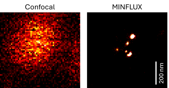
Traditional fluorescence microscopy is limited by the diffraction limit of light, which restricts resolution to about 200-300 nanometers. MINFLUX breaks this limit and allows imaging at much higher resolutions (down to a few nanometers).

MINFLUX
- Tracks labeled molecules at high frequencies up to 10 kHz, resolving molecular motion every 100 μs
- Achieves localization precisions of 1-3 nm (3D) in biological samples on large fields of view (10 x 15 μm)
- Compatible with both live and fixed fluorescently labeled specimens. Multi-color imaging is possible.

Comparison between confocal and MINFLUX resolution. Individual RNA probes hybridized along an RNA strand. © 2024 DaMBIC.
MINFLUX principle
MINFLUX works by the principle of minimal photon fluxes where fluorophore positions can be determined with a minimal number of photons and consequently within a fast and accurate spatio-temporal regime. The single emitting molecules are localized with an excitation pattern featuring a spatially well-controlled intensity zero, e.g. a donut as seen below. The intensity zero of this excitation beam is then scanned onto the sample and used to probe the fluorophore positions.

System details
- Resolution down to 1-3 nm
- The system contains two pulsed excitation laser sources @ 561 nm and @ 640 nm allowing for imaging of fluorophores in the excitation range ~550 - 680 nm.
- The MINFLUX is built onto an IX83 Olympus microscope with a multicolor CoolLED illumination source.
Specifications
Microscope body:
At DaMBIC, the MINFLUX is built on an Olympus IX83 inverted microscope with a multicolor CoolLED illumination source.
Sample holder (compatible with):
-
Glass slides, chambered slides, glass-bottom dishes (all of high-quality glass and precise thickness, #1.5H)
High-NA objective set optimized for MINFLUX.
-
100X / NA 1.45 UPlanXApo, Oil
-
40X / NA 0.9 UPlanXApo
-
20X / NA 0.8 UPlanXApo
-
2X / NA 0.1 PlanApo
Light Sources:
-
The system contains two pulsed excitation laser sources @ 561 nm and @ 640 nm allowing for imaging of fluorophores in the excitation range ~550 - 680 nm.
-
CoolLED multisource for brightfield/epifluorescence for initial localization of the sample.
Integrated single-photon-sensitive detectors for MINFLUX localization.
Stage:
-
Motorized stage (x, y)
-
Active sub-nanometer stabilization technology based on laser-illuminated fiducial markers. This keeps the sample perfectly still, with residual fluctuations < 1 nm in 3D.
Imspector (acquisition and control).
Location:
V12-605b-0