Facilitators
- Mikael Palner, Assistant Professor of Small Animal Imaging, Department of Clinical Research and Nuclear Medicine
- Peter Grupe, MD, Associate Clinical Professor, specialist in charge (Neuro Imaging), Department of Nuclear Medicine
Description
We develop methodology for functional, non-invasive and translational in vivo neuroimaging using combinations of PET, SPECT, CT and MR. These imaging modalities are unique as they work equally in both animals and humans, therefor providing absolute translational ability. In the BRIDGE network, we are mainly a support function for other disease oriented research areas, providing valuable in vivo imaging data from patients and animals, and correlate these to disease progression or functional decline.
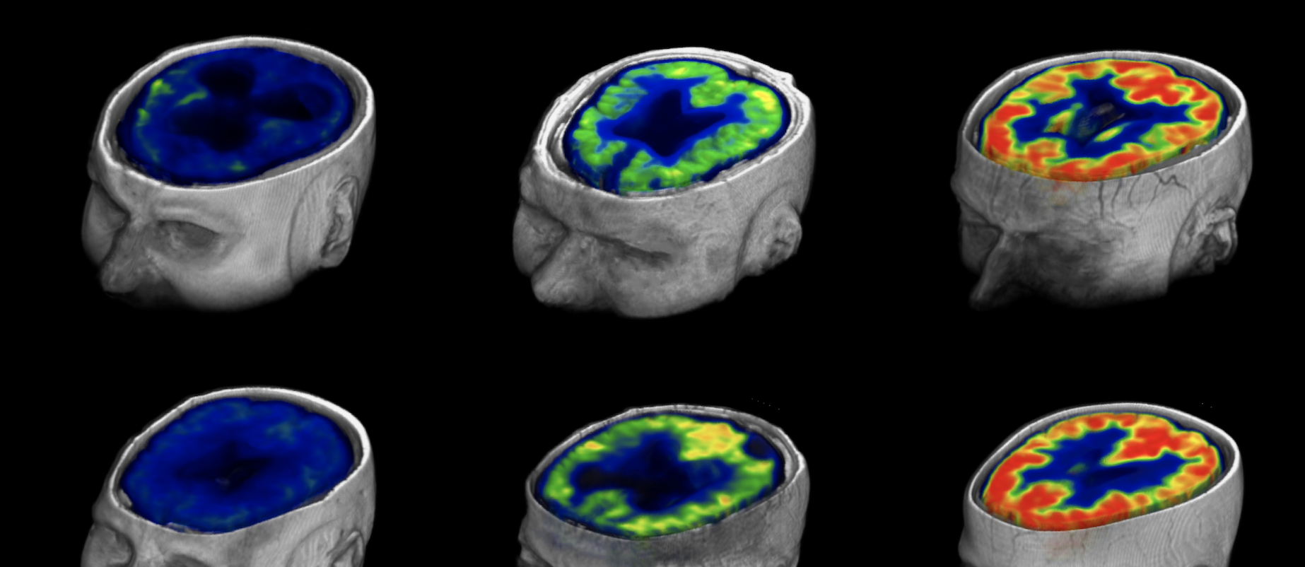
The aim of neuroimaging group is to understand brain structure and function using various advanced imaging techniques. Visualization of the brain and spinal cord, which are both inaccessible during life, has been one of the dreams of neuroscientists for many decades. This has now been achieved by techniques such as CT scan, MRI, and PET scan on daily basis which contribute tremendously to early diagnosis and treatment. However, these techniques can also serve as powerful research tools to understand brain function and structure in health and disease or discover new drugs.
Our Group consists of clinical and non-clinical researchers from the Departments of Neurology, Psychiatry, Radiology, Molecular Medicine, Nuclear Medicine, and Pathology. Together they provide extensive infrastructure and expertise to study the brain in human and animal. They try to translate basic science into diagnostic and therapeutic innovations. SDU and OUH host three 1.5T MRI, three 3T MRI, one hybrid PET/MRI and two PET traceTM 800 series cyclotrons. There is also a PET/SPECT/CT and a whole-body imaging facility for animal study. We employ various radiotracers in our PET study.
To test a new hypothesis, we may employ combined positron emission tomography (PET), magnetic resonance imaging, functional MRI, diffusion tensor imaging, and magnetic resonance spectrometry to measure brain function, structure and chemistry in volunteers or patient groups. This is now possible with the newly installed state-of-the-art PET-MR system at the Department of Nuclear Medicine.
Neuroimaging techniques allow imaging thin brain slices that are digitally put together to create 3D whole brain image. This allows study of cytoarchitecture as well as various omic studies.
If you have any questions or need support for your idea, please contact one of the above facilitators depending on your area of interest.